
Add Health Monitoring to Any Smart Device (in 3 Steps)
A concrete guide for product managers on how to add health monitoring to smart device hardware using embedded rPPG technology in three streamlined steps.
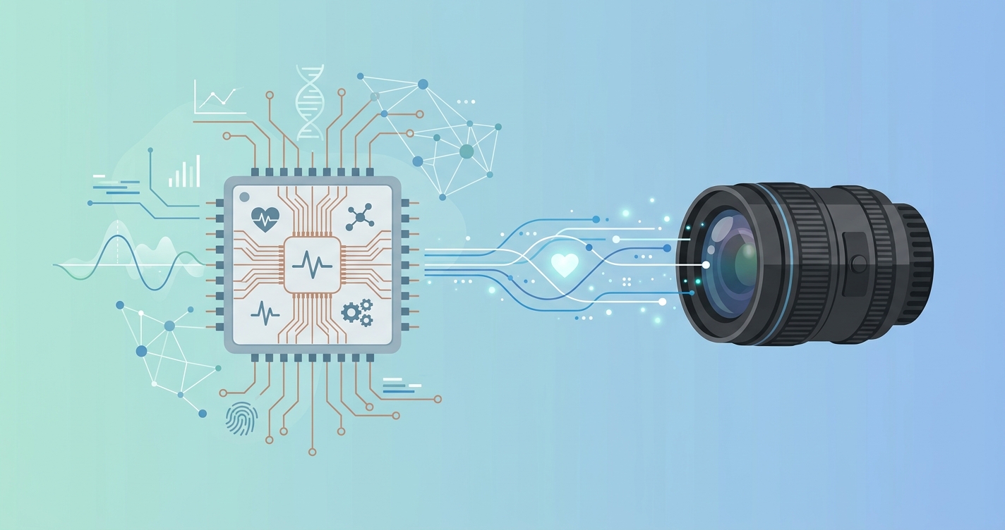
For product managers scoping the next generation of clinical hardware and connected endpoints, the bill of materials for physiological measurement is undergoing a quiet revolution. Historically, capturing a patient heartbeat or respiratory rate required dedicated physical peripherals. Product teams had to source, validate, and integrate separate hardware modules for every physiological metric they wanted to track. Now, optical sensors and advanced computer vision have transformed everyday cameras into medical grade diagnostic endpoints. The decision to add health monitoring to smart device roadmaps is no longer a question of whether it is technically feasible, but rather how quickly engineering teams can integrate the capability without inflating the hardware footprint. By moving the complexity of vital sign extraction from specialized silicon to embedded software, device manufacturers are compressing product development timelines from years to months. This transition is actively reshaping the procurement strategy for clinical kiosks, connected health tablets, and industrial safety monitors.
"The global IoT in healthcare market size was valued at USD 113.8 billion in 2023 and is expected to grow at a compound annual growth rate of 20.7 percent from 2024 to 2030, driven by the increasing adoption of connected medical devices and the growing demand for remote patient monitoring."
- Grand View Research, IoT in Healthcare Market Report, 2024
How to add health monitoring to smart device
Product teams looking to add health monitoring to smart device hardware often overestimate the friction involved in modern integrations. Because the industry spent a decade treating vital sign collection as a strictly physical hardware problem, hardware roadmaps are still burdened by legacy assumptions about procurement and clinical validation timelines. The process of embedding remote photoplethysmography (rPPG) into a clinical kiosk, patient monitoring tablet, or Internet of Things endpoint can actually be distilled into three concrete steps: hardware selection, software integration, and environmental testing.
Step 1: standardizing the hardware requirements
The transition to contactless vitals measurement means that the primary sensor is simply an RGB camera. Unlike traditional integrations that require acquiring heavily regulated blood pressure cuffs or pulse oximetry boards, embedding rPPG relies on commodity imaging components that are likely already present on the device bill of materials. The critical hardware specifications include:
- A minimum resolution of 720p at 30 frames per second to capture the micro variations in facial blood volume.
- Global shutter or well calibrated rolling shutter sensors to minimize motion artifacts when the subject moves their head.
- Adequate local compute power, typically a multi core ARM processor or a dedicated neural processing unit to handle the localized edge inference.
- An infrared sensor module if the deployment environment suffers from low ambient lighting or requires nighttime monitoring capabilities.
By standardizing around the camera module, product managers can eliminate single purpose health sensors. This reduces the total cost of manufacturing, simplifies the supply chain, and minimizes the physical footprint of the final hardware enclosure.
Step 2: integrating the embedded software engine
Once the imaging hardware is finalized, the next phase is software integration. Instead of building a computer vision pipeline from scratch, manufacturers utilize pre built software development kits to handle the complex extraction of the blood volume pulse from facial video. The mathematical complexity of filtering out ambient light noise and user motion is handled entirely by the vendor algorithm. The integration process involves:
- Initializing the rPPG engine within the host application environment and allocating the necessary memory buffers.
- Routing the raw camera frame buffer directly to the software development kit for local, frame by frame processing.
- Managing the structured data output containing real time heart rate, respiratory rate, and physiological stress metrics.
- Ensuring the user interface displays appropriate positioning feedback to the end user, such as a face bounding box or lighting warnings.
The most critical aspect of this step is ensuring that the software operates strictly on the edge. High performance embedded engines are designed to operate within the thermal and memory constraints of mobile operating systems, ensuring the device remains responsive while the health monitoring runs as a background thread.
Step 3: environmental and clinical validation testing
The final step is verifying that the integrated system performs accurately across the target deployment environments. Because remote photoplethysmography relies on ambient or structured lighting to illuminate the skin, validation testing must account for variables that affect optical clarity. Engineering teams must conduct tests across different lighting conditions, ranging from bright clinical waiting rooms with fluorescent overheads to dimly lit urgent care check in bays.
Skin tone diversity must also be validated to ensure the embedded engine processes melanin variations equitably. Furthermore, testing must cover common facial occlusions such as thick beards, reading glasses, and medical masks. Robust edge software will automatically flag when an occlusion prevents an accurate reading, allowing the user interface to prompt the patient for an adjustment rather than silently logging a false metric.
Evaluating integration paths: custom build vs embedded SDK
The decision to build an internal computer vision pipeline versus licensing a mature optical engine dictates the entire product timeline. Many hardware teams initially attempt to train open source health models, only to discover that achieving clinical accuracy in uncontrolled environments requires years of dedicated research. The following table contrasts the two approaches for device manufacturers.
| Integration Metric | Custom rPPG Development | Pre-Built SDK Integration |
|---|---|---|
| Time to Market | 18 to 24 months | 4 to 8 weeks |
| Hardware Costs | High (requires specialized validation) | Low (utilizes existing cameras) |
| Maintenance Burden | High (continuous algorithm updates) | Low (handled by vendor) |
| Core Competency | Diverts engineering resources | Keeps team focused on core product |
| Edge Performance | Often slow and unoptimized | Highly optimized for ARM/NPU processors |
Industry applications for embedded health sensors
The shift toward software defined physiological measurement is unlocking new form factors across the healthcare ecosystem. By using the three step integration process, manufacturers are deploying contactless vital signs in environments where traditional hardware was historically prohibitive due to cost, maintenance, or hygiene concerns.
Clinical kiosks and check-in stations
The modern waiting room is transitioning from a passive holding area to an active screening environment. Clinical kiosks equipped with embedded health monitoring can automatically capture a patient pulse, respiration, and baseline stress levels while the user verifies their insurance and demographic data. This parallel processing eliminates the bottleneck of manual triage and allows nursing staff to focus on direct patient care. By removing physical touchpoints like finger clips, clinics also reduce the vector for hospital acquired infections.
Iot platform endpoints
Internet of Things platform providers are extending their hardware ecosystem beyond simple environmental monitoring. By turning existing smart displays and security access panels into health screening nodes, platform providers can offer workplace wellness checks and secure facility access based on physiological baselines. This non invasive approach ensures high compliance rates without requiring users to attach physical sensors. In manufacturing environments, these endpoints can scan workers for elevated fatigue metrics before they operate heavy machinery.
Remote patient monitoring tablets
Tablet based systems are the dominant form factor for at home health monitoring. Patients recovering from surgery or managing chronic conditions often struggle with the complexity of operating multiple Bluetooth peripherals. Embedding the vitals measurement directly into the tablet camera turns the daily check in application into a frictionless, single device experience. The patient simply looks at the screen while answering their daily symptom questionnaire, and the tablet securely logs their vital signs in the background.
Current research and evidence
The transition of remote photoplethysmography from laboratory environments to commercial embedded systems is backed by extensive academic validation. Recent studies emphasize the viability of running complex physiological extraction models on localized edge hardware without sacrificing accuracy.
In 2024, researchers Rinaldi Anwar Buyung, Alhadi Bustamam, and Muhammad Remzy Syah Ramazhan at Universitas Indonesia published findings on integrating remote photoplethysmography with machine learning models. Their research demonstrated that multimodal approaches combining facial video with basic physical attributes can achieve a Mean Absolute Error of 3.057 beats per minute using optimized regression models. This level of precision confirms that edge processed optical signals can rival the accuracy of standard consumer wearables, even when deployed on modest computing hardware.
Furthermore, industry analysis confirms the financial trajectory of these integrated solutions. The 2024 Grand View Research report on the IoT in Healthcare market highlighted that the integration of artificial intelligence and machine learning is fundamentally enhancing the capabilities of local health monitoring endpoints. This research shift clearly signals an industry focus from cloud dependent analysis to localized, real time edge processing, allowing manufacturers to maintain strict patient privacy protocols while delivering immediate physiological feedback.
The future of embedded vitals
The trajectory of embedded physiological measurement points toward complete ambient monitoring. As neural processing units become standard components on even the most cost effective system on chip architectures, the computational overhead of running advanced optical extraction will approach zero. This hardware evolution will allow manufacturers to continuously monitor patient baselines in the background without requiring explicit user interaction or dedicated application states.
Future smart devices will likely feature multi modal sensor fusion, combining optical measurements with millimeter wave radar and acoustic sensors to build a comprehensive physiological profile. A clinical kiosk in 2026 will not merely report a static heart rate. It will analyze the micro fluctuations in blood volume pulse to assess cardiovascular efficiency, cross reference that data with respiratory sounds, and provide the attending physician with a holistic physiological summary before the patient even enters the examination room. This invisible integration of health tracking will ultimately make proactive healthcare a default feature of the hardware we interact with daily.
Frequently asked questions
What are the hardware limitations for adding optical sensors to a device?
The primary limitation is ambient lighting and camera quality. The device must feature a camera capable of capturing clear facial video at 30 frames per second without severe motion blur. Additionally, the local processor must have sufficient compute capacity to run the extraction algorithms without causing thermal throttling or draining the battery excessively.
Do I need to send patient video to the cloud for processing?
No. Modern embedded vitals software processes the camera feed entirely on the edge device. The facial video is analyzed in real time within the local memory buffer and immediately discarded. Only the final physiological data outputs, such as heart rate digits, are transmitted or saved, which drastically simplifies compliance with health privacy regulations.
Can an embedded camera measure continuous blood pressure?
While extracting heart rate and respiratory rate from optical signals is highly validated, absolute continuous blood pressure measurement via camera remains in the advanced research phase. Current commercial systems focus on the highly reliable extraction of the blood volume pulse, which can indicate physiological stress and cardiovascular trends, rather than diagnostic absolute blood pressure numbers.
How does skin tone affect contactless vitals integration?
Historically, optical sensors struggled with darker skin tones due to varied light absorption. Modern machine learning models are trained on highly diverse, globally representative datasets. Device manufacturers must ensure their chosen software vendor provides explicit validation data demonstrating equitable accuracy across the full Fitzpatrick skin type scale before committing to an integration.
To support product teams navigating this hardware transition, Circadify provides a highly optimized embedded rPPG engine built specifically for edge environments. The technology allows manufacturers to bypass the complexity of algorithm development and immediately test contactless vital signs on their existing hardware prototypes. To review technical documentation and request an SDK for your project, explore our Hardware integration guide.